
Peptide research papers often include pathology images, microscopy panels, and laboratory photographs. Those figures document observations for other scientists. This article explains why they appear, what kinds of questions they answer, and how non-specialist readers can interpret them cautiously. It is educational literacy about published papers, not a guide to running a lab or performing tests.
Why pathology and lab images appear in papers
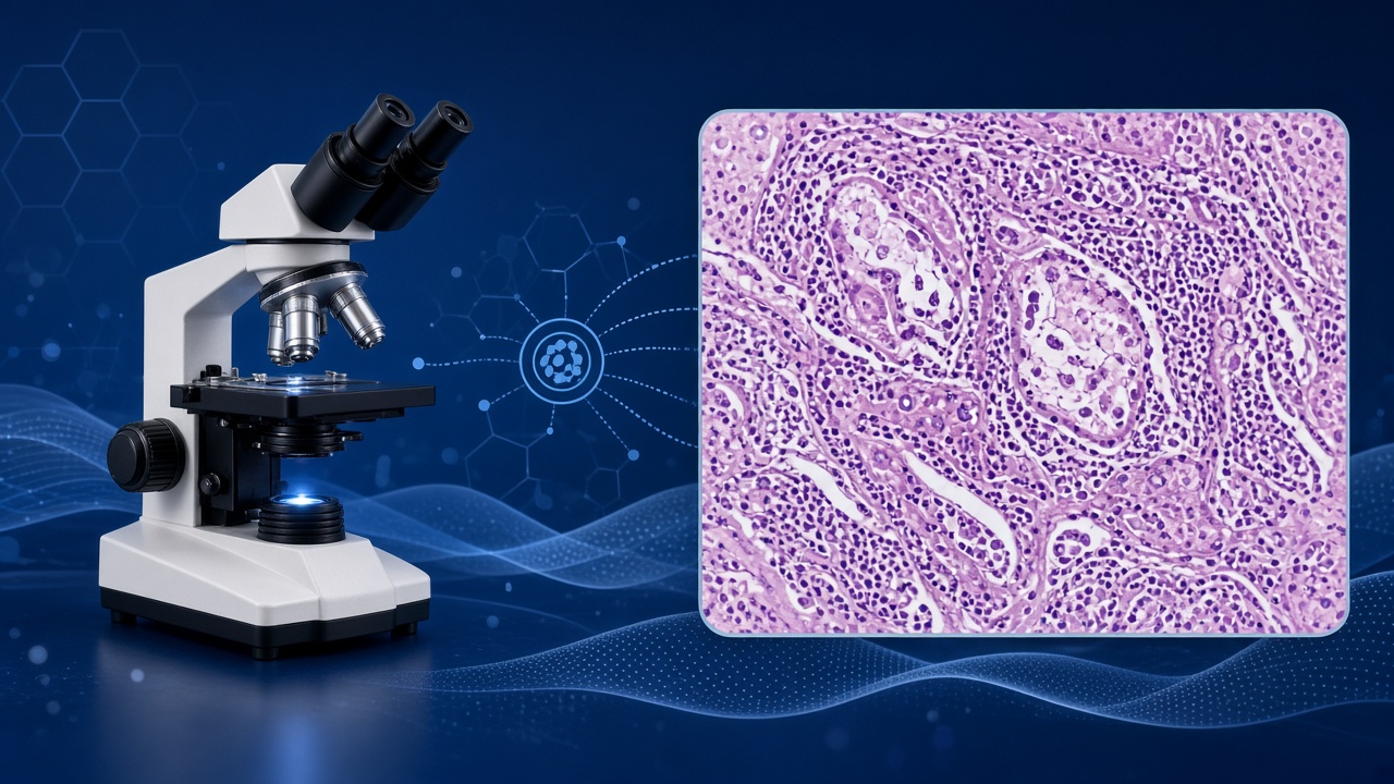
Biomedical papers are arguments supported by data. Sometimes the data are numbers. Sometimes they are images of tissue sections, cultured cells, fluorescence signals, or gross laboratory observations. Pathology-style figures help authors show what they saw under stated conditions.
In peptide research, imaging may document morphology
In peptide research, imaging may document morphology after an experimental intervention in a model system, localization of a signal, or qualitative differences between control and treated groups. The image is evidence inside a methods framework, not a standalone advertisement.
Non-specialist readers often meet these figures through social
Non-specialist readers often meet these figures through social crops that delete captions. That deletion is where misunderstanding begins. A stained section without legend can look like proof of anything a caption-writer wants.
Educational literacy restores the missing frame: what question
Educational literacy restores the missing frame: what question the figure answers, in which organism or culture system, and with which controls.
Imaging exists because some biological claims are spatial
Imaging exists because some biological claims are spatial. Location, morphology, and pattern can matter as much as a single number. That is why papers invest in figures. It is also why cropped figures without context are uniquely abusable.
Types of visual evidence readers commonly encounter
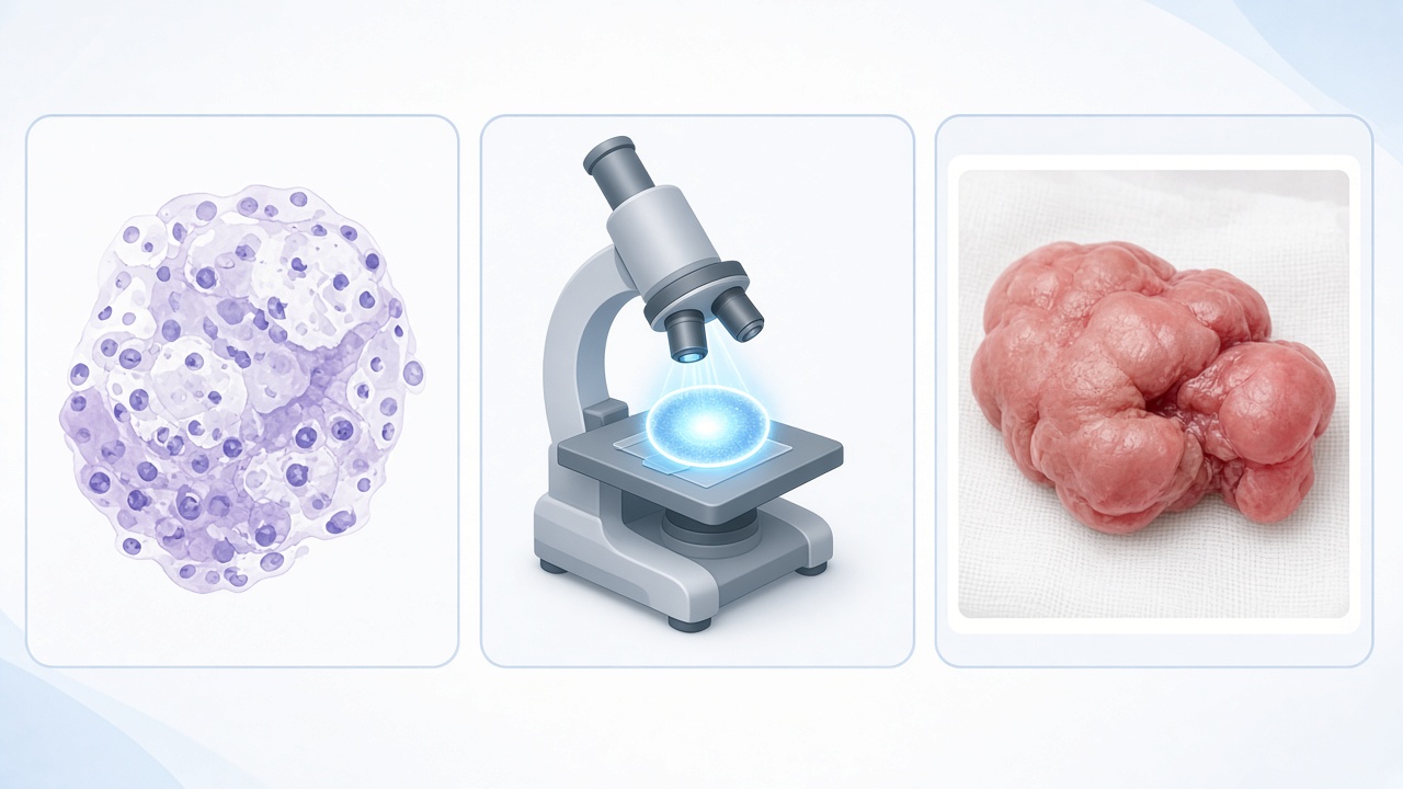
Histology panels show thin tissue sections stained to highlight structures. Microscopy panels may use light, fluorescence, or electron methods depending on the question. Gross photographs may show organs or injury models at macroscopic scale.
- Histology: tissue architecture under staining protocols described in methods.
- Fluorescence microscopy: labeled signals and localization themes.
- Gross images: macroscopic experimental observations.
- Schematics: conceptual aids, not raw observations.
- Composite panels: compare groups only after reading labels and n notes.
Visual types and claim resolution
Each visual type has a different resolution of claim. A fluorescence image might support a localization story. A histology panel might support a descriptive tissue-change story. Neither automatically supports a lifestyle outcome story.
Papers may also include schematic diagrams beside real images
Papers may also include schematic diagrams beside real images. Schematics are teaching aids. Real images are observational records. Confusing the two makes speculative cartoons feel like photographs of destiny.
When peptide topics cross into regenerative or injury-model themes
When peptide topics cross into regenerative or injury-model themes, visual evidence becomes especially persuasive and especially easy to over-read. Slow caption reading is the antidote.
Western-facing science communication sometimes treats images
Western-facing science communication sometimes treats images as universal language. They are not. A trained pathologist and a social-media audience do not see the same information density. Educational literacy narrows that gap without pretending to confer professional credentials.
Caption-first literacy
Read the caption before trusting your eyes. Captions usually state what is shown, which group is which, what the arrows mean, and what magnification or scale applies. Legends may define abbreviations that social crops remove.
Look for control panels
Look for control panels. A treated image without a control comparison is incomplete for many claim types. Look for quantification notes: sometimes authors complement images with bar graphs because qualitative impressions can mislead.
Ask whether the figure is representative and how many independent
Ask whether the figure is representative and how many independent samples support it. Good papers discuss this. Weak communication hides it.
If you cannot access the full paper
If you cannot access the full paper, be twice as cautious with isolated images. A figure without methods is a postcard, not a dataset.
Practice a caption paraphrase: rewrite the legend in your own
Practice a caption paraphrase: rewrite the legend in your own ordinary words before forming an opinion. If you cannot paraphrase it, you are not ready to circulate the image as evidence of anything.
What research images cannot do by themselves
Images cannot certify human clinical effectiveness on their own. They cannot replace safety databases, controlled trials, or regulatory review. They cannot tell a reader how to diagnose disease or how to run pathology workflows.
They also cannot settle manufacturing quality questions
They also cannot settle manufacturing quality questions for informal products. A beautiful academic figure from a controlled lab context does not validate an unrelated commercial vial discussed online.
Therapeutic peptide landscape reviews emphasize long routes
Therapeutic peptide landscape reviews emphasize long routes from experimental observations to developed medicines. Imaging can appear early in that route as mechanistic or descriptive support. Early support is not late-stage confirmation.
Keeping that ceiling visible protects readers from visual rhetoric
Keeping that ceiling visible protects readers from visual rhetoric: the sense that seeing is settling.
Images also cannot disclose conflicts
Images also cannot disclose conflicts, funding constraints, or unpublished negative replicates. Those belong to the broader paper and literature context. Figure literacy is necessary and still incomplete alone.
Common misreadings in public discussion
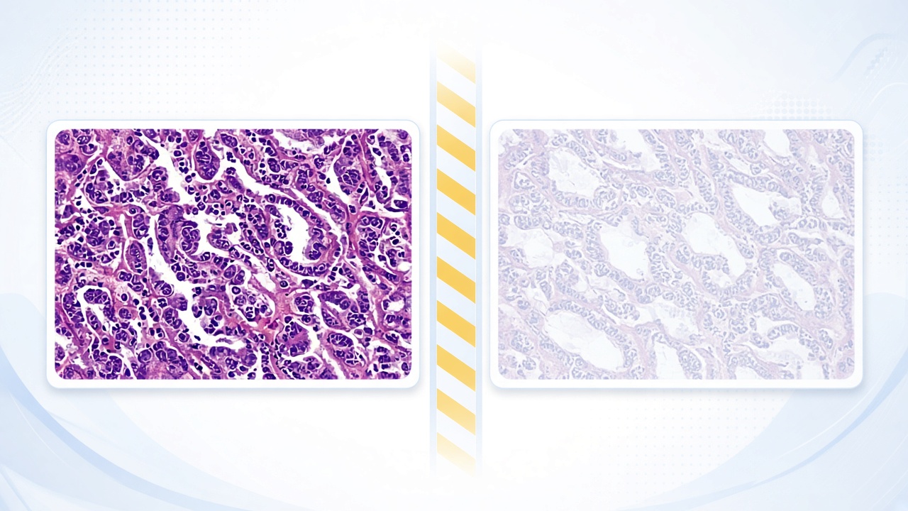
One misreading is aesthetic causation: the more dramatic the stain contrast, the stronger the assumed real-world effect. Visual drama is not effect size.
Another is species transfer: an animal histology panel is treated
Another is species transfer: an animal histology panel is treated as a human clinical photograph. Model identity matters and should stay attached to the claim.
A third is method erasure: readers ignore that staining
A third is method erasure: readers ignore that staining, sectioning, and imaging choices shape what is visible. Methods are not trivia. They are part of the evidence.
A fourth is instructional fantasy: people treat paper figures as
A fourth is instructional fantasy: people treat paper figures as if they were screenshots from a how-to course. They are not. This article will not bridge into laboratory instruction.
A fifth is selective panel shopping: choosing the most
A fifth is selective panel shopping: choosing the most extreme-looking panel while ignoring quantification that shows modest effects. Eyes prefer drama. Papers often include numbers to restrain drama. Read both.
How to practice figure literacy on peptide papers
Pick an open abstract or paper on PubMed. Scroll to a figure. Write one sentence stating only what the caption says was measured. Write a second sentence stating what you are not allowed to conclude yet. That second sentence is the educational muscle.
Compare the figure claim with the paper’s limitations paragraph
Compare the figure claim with the paper’s limitations paragraph. If social media used the figure but not the limitations, you have found the distortion point.
Use neighboring literacy pages to widen the lens
Use neighboring literacy pages to widen the lens. Assays and measurements education explains quantitative companions to images. Pharmaceutical peptide research education explains how early observations sit inside longer development contexts.
If you teach students or teammates
If you teach students or teammates, assign caption paraphrases before discussion. That single exercise prevents a surprising amount of overclaim.
Ethics of sharing research images online
Sharing a figure without caption, journal link, and limitation context spreads misunderstanding even when intentions are curious rather than malicious.
Respect publication access rules and avoid implying that a lab
Respect publication access rules and avoid implying that a lab image is medical advice for viewers. Recaptioning someone else’s figure with lifestyle promises is a communication harm.
Educational sharing looks different: full citation path
Educational sharing looks different: full citation path, model-system note, and a sentence about what remains unknown. That pattern keeps science social without making it reckless.
If a community reacts to images with protocol requests
If a community reacts to images with protocol requests, answer with literacy redirects rather than instructions. Curiosity can be welcomed while instruction stays closed.
Boundaries for this educational page

This page does not teach tissue collection, staining recipes, microscope operation, diagnostic interpretation, or laboratory safety procedures. Those are professional domains with training requirements.
It does not help readers build home labs or recreate pathology
It does not help readers build home labs or recreate pathology workflows. The slug retained from earlier site structure refers to pathology and camera-style lab documentation themes in content history, not to a consumer testing product guide.
It does not convert images from experimental peptide papers
It does not convert images from experimental peptide papers into personal health decisions. For personal decisions, use clinicians. For reading decisions, use captions and methods.
Pathology and lab imaging literacy should make papers more readable
Pathology and lab imaging literacy should make papers more readable and hype less sticky. That is the entire assignment.
It also does not evaluate clinical diagnostic imaging for patients
It also does not evaluate clinical diagnostic imaging for patients. Research figures and clinical care imaging are different contexts with different responsibilities.
Bottom line
Pathology and laboratory images appear in peptide research papers because science often needs to show what was observed, not only narrate it. Showing is powerful. Power needs caption discipline.
Read legends first, respect model systems, and refuse the jump
Read legends first, respect model systems, and refuse the jump from dramatic visuals to human protocols. Educational readers can appreciate figures without misusing them.
Continue with assays education and pharmaceutical research literacy to see how images sit beside measurements and development context. Keep Safety First Education nearby whenever content starts sounding like instruction.
Figures start evaluation, not end it
Seeing a figure is the start of evaluation, not the end of it.
If you remember one habit, remember caption-first reading. It prevents most of the avoidable mistakes this page exists to name.