
A peptide bond is the covalent link that joins amino acids into chains. Understanding that bond, how it forms, why it resists casual rotation, and how enzymes can break it, is foundational for reading peptide science. This article is educational only and is not a lab protocol or medical advice.
What a peptide bond is, in plain scientific language
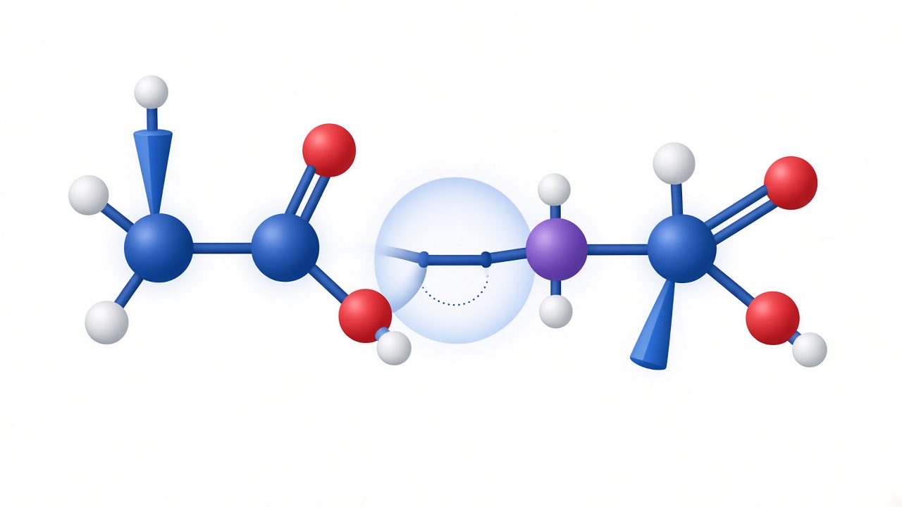
A peptide bond is the covalent connection that turns separate amino acids into a chain. IUPAC frames peptides as amides derived from two or more amino acids by forming a bond from the carbonyl carbon of one amino acid to the nitrogen of another. That definition is short, and it is enough to unlock most educational reading about peptides and proteins.
StatPearls peptide biochemistry
StatPearls material on peptides likewise starts from amino-acid polymerization through peptide bonds. Molecular Biology of the Cell chemistry chapters place the same bond at the center of how cells build macromolecules. Once you can picture the backbone repeating unit, sequence notation and residue numbering become less mysterious.
Amide bond versus peptide bond wording
The bond is often called an amide bond because the linked group resembles other amides in organic chemistry. In biomolecules, writers usually prefer peptide bond when the context is amino-acid polymers. Both phrases point to the same linkage class.
Educational scope and limits
This article stays educational. It explains concepts used in textbooks and reviews. It does not provide laboratory procedures, reagent lists, or any guidance for making or using compounds outside formal research settings.
How peptide bonds form in living cells
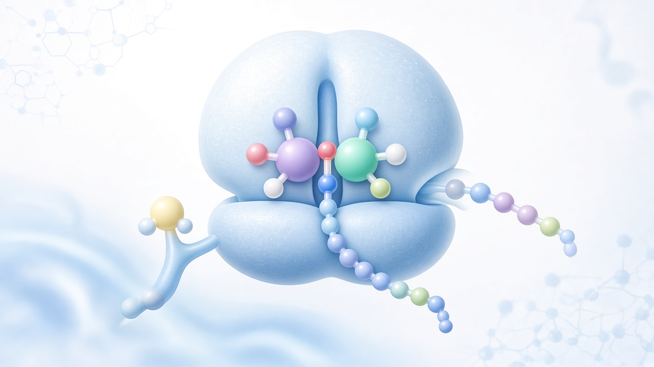
In biology, the main story of peptide-bond formation is translation. Ribosomes join amino acids in the order specified by messenger RNA. Aminoacyl-tRNA synthetases charge tRNAs, and the ribosome catalyzes peptide-bond formation as the growing chain transfers to the next amino acid. The result is a polypeptide that may later fold into a protein or be processed into shorter bioactive fragments.
Condensation picture versus cellular reality
Introductory chemistry often teaches a condensation picture: the carboxyl group of one amino acid and the amino group of another join, releasing water. That picture is useful for understanding the stoichiometry of the linkage. Cellular reality is more elaborate, because activation and catalysis are handled by the translation machinery rather than by spontaneous collision of free amino acids in water.
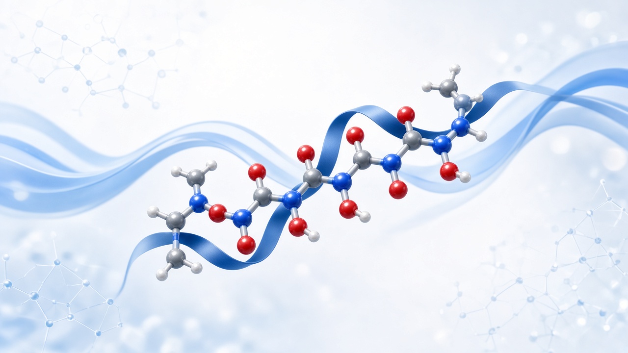
Directionality matters. Chains are described from the N-terminus to the C-terminus, reflecting how residues are ordered in the backbone. Educational figures show the repeating -N-Cα-C- pattern with side chains branching from the alpha carbons. Peptide bonds sit between the carbonyl carbon of one residue and the nitrogen of the next.
Processing after chain assembly
After synthesis, cells may cut, modify, or traffic chains. Cleavage of a precursor protein can release a peptide messenger. Those later steps still depend on the existence and accessibility of peptide bonds as chemical handles for enzymes.
Energy accounting appears in advanced texts as well. Biological peptide-bond formation is coupled to prior activation steps, which is why free amino acids in water do not spontaneously polymerize into accurate proteins. That detail protects beginners from over-simple kitchen chemistry analogies.
Planarity, resonance, and backbone geometry
Peptide bonds are not floppy single bonds in the everyday sense. Resonance delocalization between the carbonyl and the nitrogen gives the C–N linkage partial double-bond character. A major educational consequence is restricted rotation and a preference for planarity of the six atoms around the amide group.
- Partial double-bond character limits rotation about the peptide C–N bond.
- Planarity of the amide unit influences backbone conformational options.
- Trans peptide bonds predominate in folded proteins under typical conditions.
- Backbone hydrogen bonds help stabilize secondary structure in longer chains.
- Side chains decorate the backbone and tune solubility, charge, and recognition.
Planarity and secondary structure
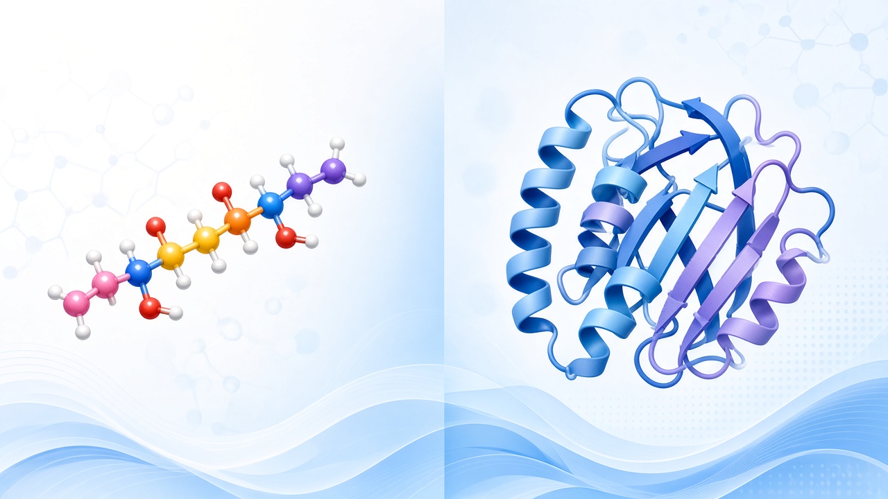
That planarity constrains backbone dihedral angles and helps explain why secondary structures such as helices and sheets are recurring themes in longer chains. Short peptides feel these constraints too, even when they do not form a full protein fold. Geometry is one reason sequence is not just a letter string; it is a three-dimensional possibility space.
Trans versus cis peptide bonds
Most peptide bonds in proteins are found in the trans configuration, which places successive alpha carbons on opposite sides of the bond. Cis peptide bonds are less common and are often discussed near proline residues in structural biology. These details appear in advanced texts, but the beginner takeaway is simpler: peptide bonds shape what folds are likely.
Backbone hydrogen bonding
Hydrogen bonding involving backbone carbonyl oxygens and amide hydrogens further organizes structure in proteins. In short peptides, similar interactions can appear locally or when a peptide binds a partner. Educational reading should separate backbone geometry facts from unsupported claims about biological outcomes.
Conformational space and crowding
Ramachandran-style thinking, even at a conceptual level, shows why some angle combinations are crowded while others recur in helices and sheets. You do not need to memorize plots to benefit from the idea that peptide-bond geometry narrows conformational space.
How peptide bonds are broken: hydrolysis and proteases
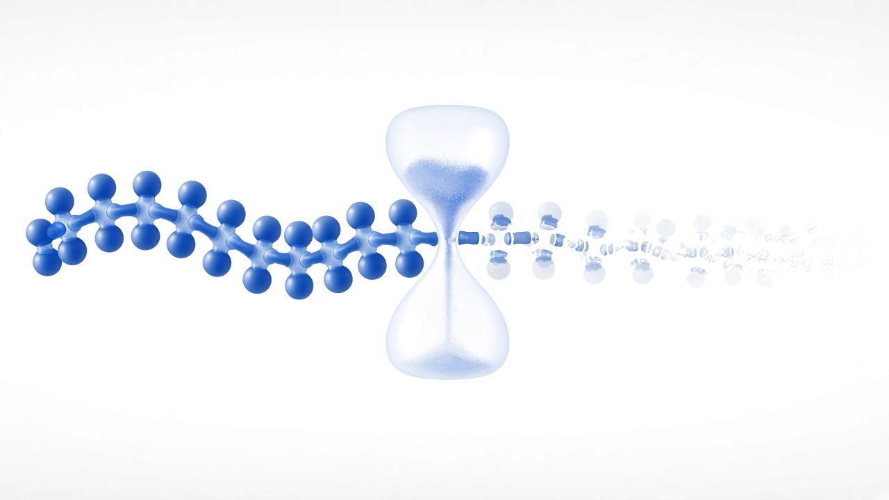
Forming a peptide bond builds a chain; breaking a peptide bond shortens or dismantles it. The chemical reverse of condensation is hydrolysis: water is used to split the amide linkage back toward carboxyl and amino components. In water alone, peptide bonds are relatively resistant to casual hydrolysis on biological timescales, which is part of why proteins can be durable information-bearing polymers.
Proteases and controlled cleavage
Cells therefore rely on enzymes. Proteases catalyze peptide-bond cleavage with specificity patterns that depend on active-site chemistry and local sequence. Digestion, blood clotting cascades, apoptosis pathways, and protein quality control all use controlled proteolysis as a theme. Educational peptide half-life discussions often return to protease vulnerability of short chains in experimental systems.
Processing cleavage as a biological feature
Not every cleavage is destructive waste. Processing enzymes can convert inactive precursors into active messengers by cutting at defined sites. In that sense, breaking selected peptide bonds is a biological feature, not only a failure mode.
Teaching hydrolysis is not a protocol
Harsh non-biological conditions can also hydrolyze peptide bonds, which is why chemistry textbooks mention acid or base catalyzed hydrolysis in teaching contexts. That teaching point is not a protocol. On this site, synthesis methodology articles remain high-level comparisons of research approaches, not step-by-step lab instructions.
Protease specificity and stability
Specificity language in protease papers, such as preferred residues around a cut site, is another literacy marker. It explains why two peptides of similar length can differ in experimental stability without invoking mystical explanations.
Why peptide-bond literacy helps when reading research
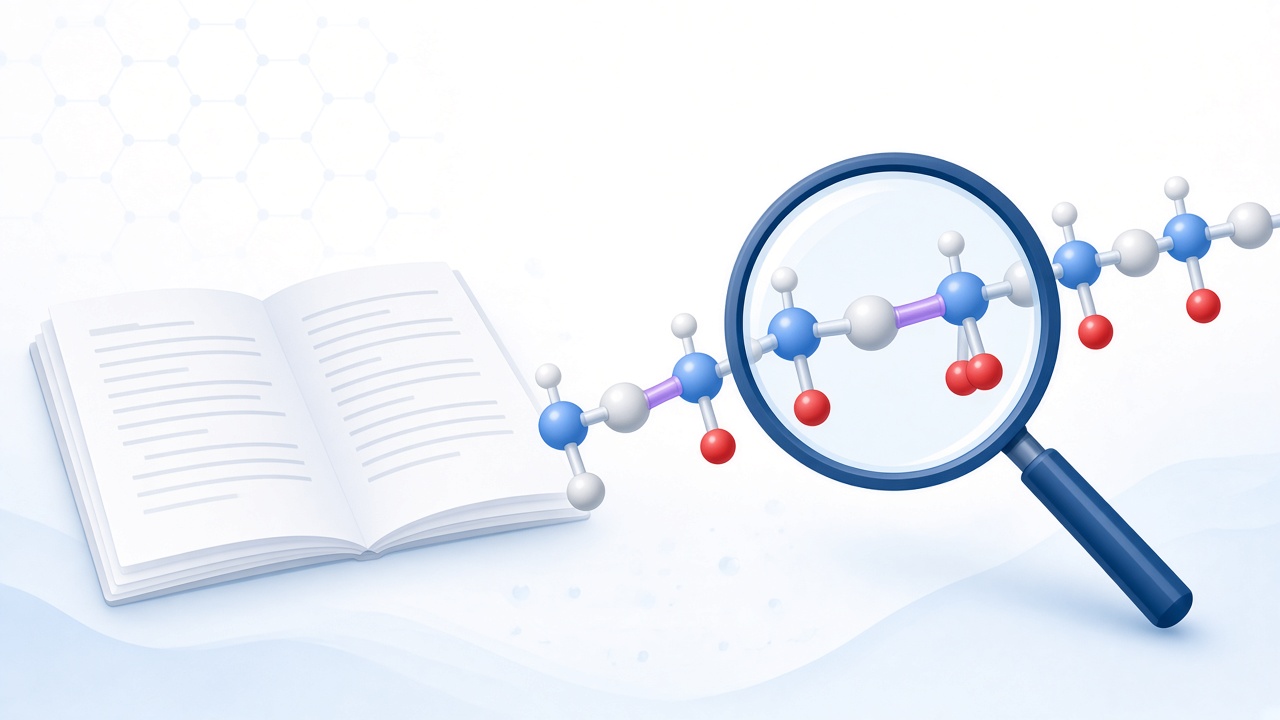
When papers discuss stability, they are often talking about whether peptide bonds remain intact in a given environment. When papers discuss receptor recognition, they may still depend on a backbone geometry that presents side chains in a useful pattern. When papers discuss fragments, they are usually describing products of selective bond cleavage from larger proteins.
Peptidomimetics after the basic bond
Readers also encounter modified backbones in advanced literature, such as peptidomimetics designed to resist proteolysis. Those topics sit downstream of the basic amide-bond idea. Learn the standard peptide bond first, then interpret modifications as deliberate changes to geometry, hydrogen bonding, or enzyme recognition.
Keep claims proportional
Keep claims proportional. A clear explanation of peptide bonds does not justify personal experimentation. It supports better reading of abstracts, figures, and educational reviews.
Common misconceptions about peptide bonds
One misconception is that peptide bonds are fragile everywhere. In reality, they are chemically sturdy enough to carry biological information, yet enzyme-accessible enough for regulated turnover. Stability is context dependent.
Another common misconception
Another misconception is that any discussion of bond formation is implicitly an invitation to synthesize compounds at home. Educational chemistry describes nature and research concepts. It does not authorize unsafe or unlawful activity, and this site does not provide protocols.
A third common misconception
A third misconception is that understanding the bond explains every headline about research peptides. Bond chemistry is necessary background, not a sufficient basis for efficacy or safety conclusions about specific experimental sequences.
A fourth common misconception
A fourth misconception is that peptide bonds only matter inside exotic research topics. In reality, the same linkage appears in ordinary nutrition and cell biology teaching. Learning the bond is general science literacy first, research-headline literacy second.
Layered literacy habit
A better habit is layered literacy: bond definition, length and folding categories, evidence type in papers, and clear separation of education from advice. That habit keeps curiosity productive.
Backbone notation readers see in papers and textbooks
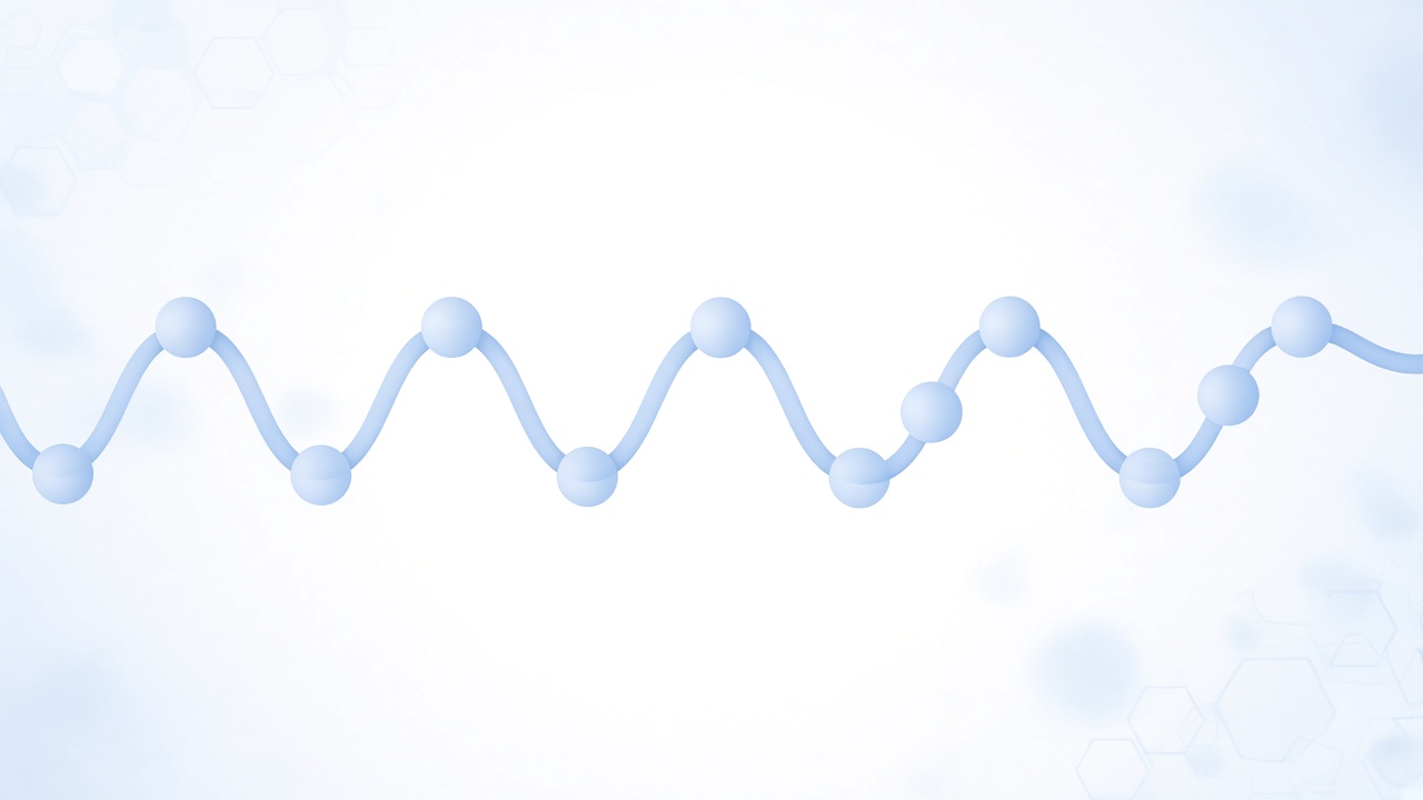
Sequence strings written in single-letter or three-letter amino-acid codes imply an ordered backbone of peptide bonds even when the bonds are not drawn. When a figure shows sticks or ribbons, the continuous trace still depends on those linkages. Learning to see the invisible bonds in a letter string is a practical reading skill.
N-terminal and C-terminal labels
Termini labels also depend on peptide-bond orientation. N-terminal and C-terminal language tells you which end has a free amino group character and which end has a free carboxyl group character in the unmodified linear case. Modifications can cap termini, and educational texts usually flag those caps because they change recognition and stability discussions.
Disulfides versus peptide bonds
Disulfide bonds and other cross-links are sometimes confused with peptide bonds by beginners. Disulfides link side chains of cysteine residues; peptide bonds link the backbone. Both can stabilize structure, but they are different chemical stories. Keeping them separate prevents misreading methods and figure legends.
Constraints build on the standard bond
When reviews discuss constrained peptides, they may combine backbone amide geometry with extra cycles or bridges. The starting point remains the standard peptide bond. Constraints are additions to that foundation, not replacements for it.
Where to go next after learning peptide bonds
If this bond explainer clarified the backbone, revisit What Are Peptides for classification language, then Peptides vs Proteins for the length and function contrast. For manufacturing concepts as discussed in literature, read Peptide Synthesis Methodologies as an educational overview only.
Primary educational source anchors
Primary educational sources cited here, IUPAC terminology, StatPearls peptide biochemistry, and Molecular Biology of the Cell chemistry chapters, are good external anchors when you want to verify definitions beyond any single website.
Remember the framing
Remember the framing: peptide bonds join amino acids into the chains that biology and research discuss as peptides and proteins. Knowing the bond helps you read those discussions accurately.
A practical reading checklist
If you want a compact study checklist, define the bond, sketch N-to-C direction, explain planarity in one sentence, and explain protease cleavage as catalyzed hydrolysis. Those four points cover most beginner needs before compound-specific pages.